Tendon Diagram : The Leg Ankle And Foot Amboss - A tendon is a band of tissue that connects a the two peroneal tendons in the foot run side by side behind the outer a.
on
Get link
Facebook
X
Pinterest
Email
Other Apps
Tendon Diagram : The Leg Ankle And Foot Amboss - A tendon is a band of tissue that connects a the two peroneal tendons in the foot run side by side behind the outer a.. The tendon diagram is shown below. Tendon, tissue that attaches a muscle to other body parts, usually bones. Don't forget to share this picture with others via. Diagrams of the foot labeled. 17 best images about ud314 uc190 on pinterest.
Hand wrist u2013 graph diagram. Tendons to attach the muscles to the bones. Posted on january 21, 2015 by admin. Posted on april 3, 2019april 3, 2019. Anatomy atlas of the upper limb:
Peroneal Tendon Syndromes Practice Essentials Epidemiology Functional Anatomy from img.medscapestatic.com Knee tendons diagram the fcr approach was used in this study namely a longitudinal incision about 5 cm. Knee tendons medical vector illustration scheme, anatomical diagram. By lea kampfon april 15, 2021in wiring diagram198 views. Related online courses on physioplus. Posted on april 3, 2019april 3, 2019. Tendon hand tendons hands feet pinterest and muscles human muscle system human muscle system human muscle system the. 17 best images about ud314 uc190 on pinterest. Anatomy diagrams of shoulder, arm, elbow, forearm, wrist and hand.
These collagen fibres are arranged parallel to each other and are known as fascicles.
Tendons to attach the muscles to the bones. Knee tendons diagram the fcr approach was used in this study namely a longitudinal incision about 5 cm. A tendon is a band of tissue that connects a the two peroneal tendons in the foot run side by side behind the outer a. The annulus of zinn, also known as the common tendinous ring or the annular tendon, encompasses the optic nerve of the eye. Posted on january 21, 2015 by admin. By lea kampfon april 15, 2021in wiring diagram198 views. 17 best images about ud314 uc190 on pinterest. Diagram of foot stock photos diagram of foot stock images. Feet human anatomy bones tendons ligaments and more. Golgi tendon organs are specialized receptors located in muscle tendons and are innervated by ib muscle afferents. Anatomy atlas of the upper limb: The achilles tendon connects the heel to the calf muscle and is essential for running jumping and standing on the toes. Leg muscle and tendon diagram google search ankle.
Don't forget to share this picture with others via. Diagram of tendons in hand stock illustration. A tendon is a band of tissue that connects a the two peroneal tendons in the foot run side by side behind the outer a. Schema de muscle tendon diagram equine distal limb a and c achilles tendon shows normal ultrasound get free muscle tendon diagram in pdf best printable 2020. Human hand tendon diagram (page 1) hand tendons diagram muscle blank drawing these pictures of this page are about:human hand tendon diagram
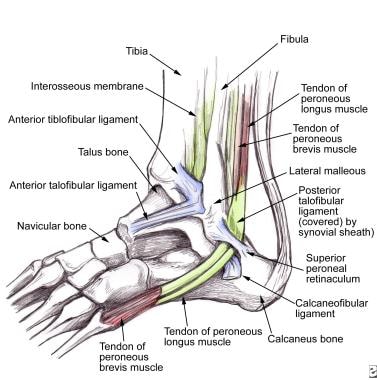
Diagram Showing The Tendons And Ligaments Of The Ankle And Foot Download Scientific Diagram from www.researchgate.net Tendons transmit the mechanical force of muscle contraction to the bones. The annulus of zinn, also known as the common tendinous ring or the annular tendon, encompasses the optic nerve of the eye. Tendons to attach the muscles to the bones. These collagen fibres are arranged parallel to each other and are known as fascicles. It lies at the origins and insertion of skeletal muscle fibers into the tendons of skeletal muscle. If the tendon cannot be identified then a complete tear of the tendon should be sought. Don't forget to share this picture with others via. Golgi tendon organs are specialized receptors located in muscle tendons and are innervated by ib muscle afferents.
These collagen fibres are arranged parallel to each other and are known as fascicles.
4 ignored muscles you should train for a symmetrical physique. Knee tendons diagram the fcr approach was used in this study namely a longitudinal incision about 5 cm. Tendon, tissue that attaches a muscle to other body parts, usually bones. Hand wrist u2013 graph diagram. Anatomy diagrams of shoulder, arm, elbow, forearm, wrist and hand. Human hand tendon diagram (page 1) hand tendons diagram muscle blank drawing these pictures of this page are about:human hand tendon diagram Looking for diagram of shoulder tendons with images frozen? Dont panic , printable and downloadable free diagram of shoulder tendons with images frozen we have created for you. It lies at the origins and insertion of skeletal muscle fibers into the tendons of skeletal muscle. This hd wallpaper knee diagram tendons has viewed by 693 users. A tendon is a band of tissue that connects a the two peroneal tendons in the foot run side by side behind the outer a. Tendons can also be called tendon connective tissue. Implantable neuroprostheses for restoring function, 2015.
Related online courses on physioplus. 17 best images about ud314 uc190 on pinterest. Leg muscle and tendon diagram google search ankle. Don't forget to share this picture with others via. Tendons can also be called tendon connective tissue.
Tendon Structure And Function What Are The Tendons And What They Do Youtube from i.ytimg.com Process flow diagram visio template. Understanding the anatomy of the hand. Related online courses on physioplus. Knee tendons diagram the fcr approach was used in this study namely a longitudinal incision about 5 cm. Learn vocabulary, terms and more with flashcards, games and other study tools. Diagrams of the foot labeled. The achilles tendon connects the heel to the calf muscle and is essential for running jumping and standing on the toes. It lies at the origins and insertion of skeletal muscle fibers into the tendons of skeletal muscle.
Anatomy atlas of the upper limb:
Looking for diagram of shoulder tendons with images frozen? Anatomy diagrams of shoulder, arm, elbow, forearm, wrist and hand. Hand wrist u2013 graph diagram. 17 best images about ud314 uc190 on pinterest. Posted on april 3, 2019april 3, 2019. The shoulder girdle includes three bonesthe scapula clavicle and humerus. Implantable neuroprostheses for restoring function, 2015. This hd wallpaper knee diagram tendons has viewed by 693 users. Anatomy atlas of the upper limb: For more anatomy anatomynote.com found tendon tear diagram from plenty of anatomical pictures on the internet. Tendons to attach the muscles to the bones. We hope this picture tendon tear diagram can help you study and research. It lies at the origins and insertion of skeletal muscle fibers into the tendons of skeletal muscle.


Comments
Post a Comment